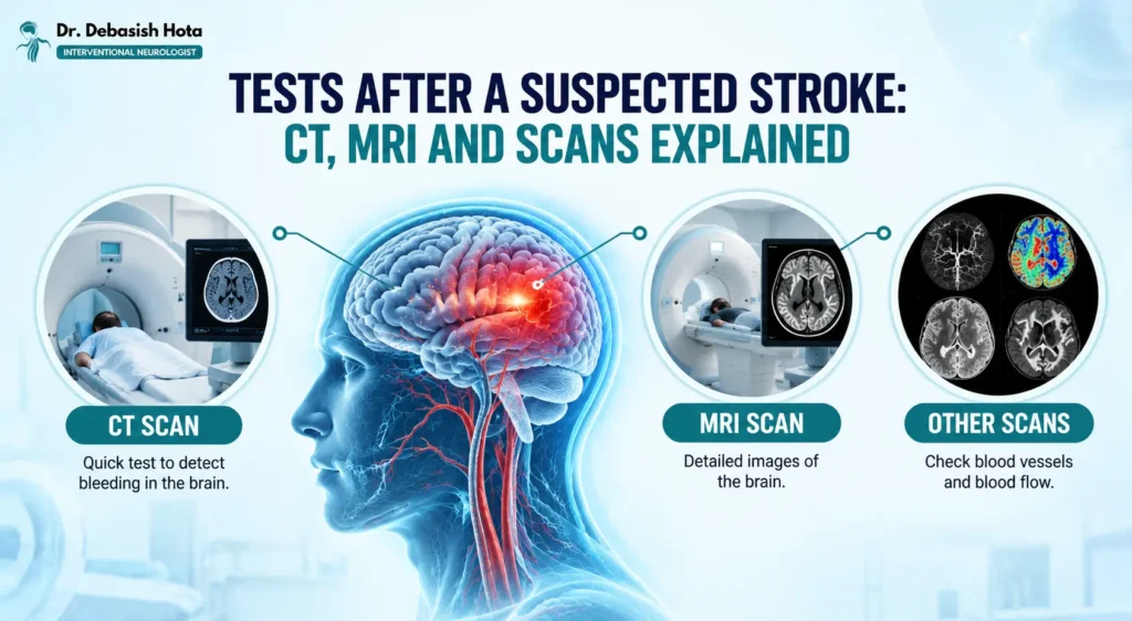
When someone shows signs of a stroke, the hospital does not wait. Within minutes, a series of tests begins to answer three urgent questions: Is it really a stroke? Is it caused by a clot or by bleeding? And which treatment can be given right now?
In this blog, we explain the tests done after a suspected stroke, what each scan shows and why the order of these tests matters.
Why Are Tests So Important in a Suspected Stroke?
Stroke symptoms can sometimes resemble other conditions, including very low blood sugar, seizures or migraine. Doctors assess the symptoms, perform a neurological examination, check blood glucose and arrange urgent brain imaging to help identify the cause.
Brain imaging is particularly important because an ischemic stroke, caused by a blocked blood vessel, and a haemorrhagic stroke, caused by bleeding, require different treatment approaches. A non-contrast CT scan is commonly used first to look for bleeding. Further imaging and tests may be needed to determine the type of stroke, assess affected blood vessels and guide treatment.
Step 1: Quick Neurological Examination
Before any scan, the doctor checks the patient’s alertness, speech, vision, face, arm and leg strength, balance and coordination. Doctors often use a scoring tool called the NIHSS (National Institutes of Health Stroke Scale) to measure how severe the stroke is.
This takes only a few minutes and happens alongside basic checks of blood pressure, heart rate, oxygen level and blood sugar.
Step 2: CT Scan of the Brain (Non-Contrast CT)
Why it matters: A non-contrast CT scan helps doctors identify bleeding in the brain and assess whether urgent stroke treatment may be appropriate. However, a normal early CT does not rule out an ischemic stroke. If bleeding is not seen, doctors consider the patient’s symptoms, when they began or when the patient was last known to be well, relevant medical history and other eligibility criteria before deciding on treatment.
Step 3: CT Angiography (CTA)
A CT angiography uses a contrast dye injected through a vein to show the blood vessels of the brain and neck.
What it shows:
- The exact location of a blocked artery
- Narrowing in the carotid arteries of the neck
- Large vessel blockages that may need a clot-removal procedure
- Aneurysms or abnormal vessels in cases of bleeding
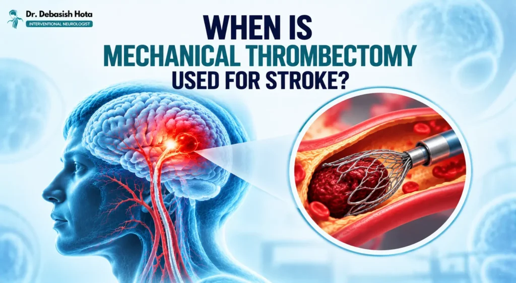
This scan helps decide whether the patient needs mechanical thrombectomy, a procedure to remove the clot directly from the artery.
Step 4: CT Perfusion Scan
A CT perfusion scan shows how well blood is flowing through different parts of the brain.
What it shows:
- The part of the brain that is already badly damaged (the core)
- The part that is still at risk but can be saved (the penumbra)
This information is especially useful when a patient arrives late. Even many hours after symptoms begin, some patients may still benefit from treatment if enough brain tissue can be saved.
Step 5: MRI Scan of the Brain
An MRI scan uses a strong magnetic field to produce very detailed images of the brain, and it is more sensitive than CT for detecting a recent stroke.
What it shows:
- Very early damage from an ischemic stroke, often within minutes (using a sequence called DWI)
- Small strokes that a CT may miss
- Strokes in the brainstem and the back of the brain, which are hard to see on CT
- The age and extent of the stroke
MRI also helps explain other brain problems. For example, repeated small strokes seen on MRI can be a cause of vascular dementia, which is why doctors sometimes order it for memory problems after a stroke.
Note: MRI takes longer than CT and may not be suitable for patients with certain metal implants or pacemakers, so doctors choose the right scan for each patient.
Step 6: MR Angiography (MRA)
An MR angiography is an MRI-based scan of the brain’s blood vessels. It can be done with or without contrast dye. It helps find narrowed or blocked arteries and is useful when a CT angiogram is not suitable.
Step 7: Carotid Doppler Ultrasound
The carotid arteries on both sides of the neck carry blood to the brain. A carotid Doppler is a painless ultrasound test that checks these arteries for narrowing or plaque.
What it shows:
- Blockage or narrowing in the neck arteries
- How fast blood is flowing through them
This test helps doctors decide how to prevent another stroke.
Step 8: Cerebral Angiography (DSA)
A digital subtraction angiography (DSA) is the most detailed test of the brain’s blood vessels. A thin tube (catheter) is passed through an artery, usually from the wrist or groin, up to the brain, and contrast dye is injected to show the vessels in real time.
What it shows:
- Exact blockages, aneurysms and abnormal vessel tangles
- Fine details of blood flow that other scans may miss
DSA is more invasive than other tests, so it is used when other scans cannot give a clear answer or when treatment is planned. It is also the platform used for procedures in neurointervention, such as clot removal and aneurysm treatment, so diagnosis and treatment can often be done in the same session.
Step 9: Heart Tests (ECG, Echocardiography and Heart Rhythm Monitoring)
Some ischemic strokes occur when a blood clot forms in the heart and travels to the brain. Heart investigations can help doctors identify possible sources of a clot or an abnormal heart rhythm and guide measures to reduce the risk of another stroke.
Common tests include an ECG to check heart rhythm, echocardiography to examine the heart’s structure and function, and longer-term rhythm monitoring when an intermittent abnormal heartbeat is suspected. The tests recommended depend on the patient’s history and initial findings.
Step 10: Blood Tests
Blood tests are done quickly and often alongside the scan. They check:
- Blood sugar: Very low sugar can mimic a stroke, and high sugar can worsen outcomes
- Complete blood count: To detect anaemia, infection or low platelets
- Clotting tests (PT/INR, aPTT): To check how well the blood clots before giving treatment
- Cholesterol (lipid profile): To assess risk of future strokes
- Kidney function and electrolytes: To make sure contrast dye and medicines are safe
- HbA1c: To check long-term diabetes control
- Cardiac markers: To check for a heart attack, which can occur alongside a stroke
Which Test Is Done First? A Quick Summary

| Test | What It Mainly Shows | When It Is Used |
| Neurological exam and blood sugar | Severity of stroke, stroke mimics | Immediately |
| Non-contrast CT | Bleeding in the brain | First scan |
| CT angiography | Blocked or narrowed vessels | Right after CT |
| CT perfusion | Salvageable brain tissue | Selected patients |
| MRI (DWI) | Early or small strokes | When CT is unclear |
| MRA | Brain blood vessels without radiation | Selected patients |
| Carotid Doppler | Neck artery narrowing | Within the first days |
| DSA | Detailed vessel map, treatment planning | Selected patients |
| ECG, Echo, Holter | Heart cause of stroke | During admission |
| Blood tests | Safety and risk factors | Immediately |
How Quickly Are These Tests Done?
Stroke assessment and brain imaging are performed urgently because treatment options may depend on how much time has passed since symptoms began. Many stroke systems aim to obtain the initial brain scan within approximately 20 minutes of hospital arrival for eligible patients, although actual timings vary by hospital and circumstances.
Some patients may be eligible for clot-dissolving treatment within a limited time window, while selected patients may benefit from mechanical thrombectomy later, based on clinical assessment and imaging findings. If you notice sudden facial drooping, arm weakness or difficulty speaking, call 112 in India or your local emergency number immediately. Do not wait for symptoms to improve or drive yourself to hospital.
What Happens After the Tests?
Once the type and cause of stroke are clear, treatment begins right away. This may include clot-dissolving medicines, thrombectomy, blood pressure control, management of bleeding or early rehabilitation. The test results also guide long-term prevention, such as medicines for blood pressure, cholesterol and blood thinning.
For a complete understanding of how the condition is treated, you can explore the details of stroke care in Bhubaneswar.
When Should You Consult a Neurologist?
Sudden facial drooping, arm weakness, difficulty speaking or other possible stroke symptoms require immediate emergency medical attention. Do not wait for a routine appointment, even if the symptoms improve.
After emergency assessment, a neurologist can help interpret scan reports, investigate possible causes of stroke and discuss appropriate treatment and prevention plans.
Dr. Debasish Hota is a Consultant Interventional Neurologist in Bhubaneswar. For non-emergency follow-up or guidance on previous scan reports, visit the doctor’s website or contact the clinic to enquire about an appointment.
Final Thoughts
After a suspected stroke, tests such as CT, MRI, angiography, heart tests and blood work work together to confirm the diagnosis, find the cause and guide life-saving treatment. The faster these tests are done, the more brain can be saved. Recognising symptoms early and reaching a stroke-ready hospital without delay makes the biggest difference.
Disclaimer: This blog is for general awareness only and does not replace professional medical advice. If you or someone near you shows signs of a stroke, seek emergency medical care immediately.
FAQs :-
What is the first test done for a suspected stroke?
A non-contrast CT scan of the brain is usually the first test. It is quick and shows whether there is bleeding in the brain, which decides the next treatment step. A blood sugar check is also done immediately, since low sugar can look like a stroke.
Is MRI or CT better for diagnosing a stroke?
Both have a role. CT is faster and is preferred in emergencies to rule out bleeding. MRI is more sensitive for detecting early and small strokes, especially in the back of the brain. Doctors often use CT first and MRI when more detail is needed.
Can a CT scan miss a stroke?
Yes. In the first hours, a CT scan may look normal even when an ischemic stroke has begun, especially for small strokes or those in the brainstem. This is why doctors combine CT with angiography or an MRI, and rely on symptoms and examination as well.
How long does it take to diagnose a stroke?
In a well-equipped hospital, the first scan is completed within about 20 to 25 minutes of arrival. Further tests such as angiography, heart tests and blood work may continue over the following hours and days to find the exact cause.
What blood tests are done after a stroke?
Doctors check blood sugar, a complete blood count, clotting tests, cholesterol, kidney function, electrolytes and HbA1c. These help confirm that treatment is safe and identify risk factors that need long-term control.